Polymer Micelles for Bioimaging: Design Strategies, Probe Loading, and Imaging Performance
Polymer micelles have become increasingly valuable in bioimaging research because they do more than passively carry contrast agents or fluorescent probes. As self-assembled nanostructures built from amphiphilic polymers, they provide a controllable microenvironment for organizing imaging units, stabilizing hydrophobic or aggregation-prone probes, and modulating how signals are generated, retained, or activated in aqueous and biologically relevant conditions. Their core-shell architecture makes them especially useful when imaging performance depends not only on the probe itself, but also on probe location, colloidal stability, background suppression, and the ability to connect signal generation with structural change. For this reason, polymer micelles are now widely considered as imaging platforms rather than simple carriers, particularly in systems involving fluorescence, near-infrared imaging, magnetic resonance-related probes, responsive imaging, and multimodal designs. This page focuses on how polymer micelles support bioimaging through probe loading, signal design, responsive activation, and multimodal integration from a polymer materials perspective.
Resources
In bioimaging research, polymer micelles are best understood as self-assembled nanoscale platforms that connect amphiphilic polymer design with imaging function. When block copolymers organize in aqueous media, they form a hydrophobic interior and a hydrated outer corona, creating a compartmentalized structure that can host imaging probes, protect sensitive imaging units, or display functional motifs at the interface. Their relevance lies not only in nanoscale transport, but also in the way self-assembly affects signal output, probe distribution, and environmental responsiveness. As a result, polymer micelles for bioimaging should be treated as structure-signal systems in which colloidal behavior and imaging performance are designed together from the beginning.
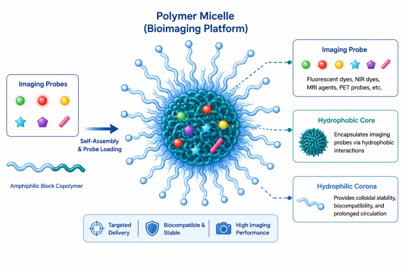 Fig. 1. Polymer micelles organize imaging probes for bioimaging applications (BOC Sciences Authorized).
A polymer micelle becomes an imaging platform when its self-assembled structure contributes directly to the behavior of the imaging unit. The hydrophobic core can solubilize or confine poorly water-compatible probes, while the hydrophilic shell helps maintain dispersion and reduces uncontrolled aggregation in aqueous environments. Beyond that, the micellar interface can regulate local polarity, molecular packing, rotational freedom, and probe-probe distance, all of which can influence fluorescence intensity, quenching, relaxivity behavior, or other signal properties. In other words, the micelle does not merely contain an imaging probe; it changes the probe's functional context.
Why Bioimaging Requires Different Micelle Design Priorities
Imaging-oriented micelles are not designed under the same priorities as drug delivery-oriented micelles. In bioimaging, high loading is not always the main objective. Instead, designers often prioritize signal clarity, suppression of background emission, preservation of probe stability, and the ability to correlate imaging changes with structural or environmental changes. A micelle that carries a large amount of probe but causes severe self-quenching or rapid leakage may be poor for imaging even if it looks acceptable as a delivery carrier. Imaging performance therefore depends on balancing structural stability with optical or magnetic functionality in a way specific to the chosen modality.
Polymer Micelles vs Other Nanocarriers for Imaging Applications
Compared with broader polymer nanoparticle systems, polymer micelles are especially attractive when reversible self-assembly, core-shell compartmentalization, and nanoscale probe organization are central to the design logic. More rigid nanoparticles may provide stronger matrix retention, while inorganic systems may offer distinctive physical contrast mechanisms. However, polymer micelles often provide a more adaptable platform when the goal is to control probe microenvironment, tune signal activation, or integrate responsive disassembly with imaging readout. Their value is therefore highest when signal behavior depends strongly on local molecular arrangement rather than on simple bulk encapsulation.
Why Signal Function and Colloidal Structure Must Be Designed Together
A polymer micelle can only function as a reliable imaging system if the colloidal structure and the signal mechanism are mutually compatible. Changes in micelle size, packing density, corona hydration, or core mobility can all influence probe behavior. Likewise, introducing a highly conjugated dye, magnetic unit, or multimodal probe may change the assembly process itself. For this reason, polymer micelle bioimaging design cannot be reduced to "select a probe and then encapsulate it." The material architecture must be planned so that signal generation remains interpretable and stable within the assembled colloidal framework.
Why Polymer Micelles Are Useful for Bioimaging?
Polymer micelles are useful for bioimaging because they address multiple limitations that free probes often face in aqueous and biologically relevant environments. Many imaging units suffer from poor water compatibility, rapid aggregation, signal instability, or uncontrolled interactions with surrounding components. A well-designed micelle can improve these conditions by organizing the probe in a more controlled nanoscale environment. Just as importantly, the micelle can influence where the probe resides, when the signal becomes active, and how readily background interference is suppressed.
Encapsulation and Protection of Imaging Probes
Many optical and hybrid imaging agents are sensitive to surrounding solvent, aggregation, oxidation, or interfacial instability. Encapsulation within a micelle core can reduce direct exposure to aqueous media and help maintain the functional state of the probe. This is particularly relevant for hydrophobic fluorophores, aggregation-sensitive chromophores, and certain conjugated signal units. In some cases, the micelle also reduces the risk of premature interaction with competing molecules in solution, allowing the imaging agent to remain in a more controlled chemical environment until measurement or activation occurs.
Improved Aqueous Dispersion and Probe Stability
Free imaging probes frequently exhibit poor dispersion in water or form irregular aggregates that generate unstable or misleading signals. The hydrophilic corona of a micelle can improve aqueous compatibility and maintain a more consistent colloidal state. This improves reproducibility and helps distinguish intended signal behavior from artifacts caused by precipitation or broad aggregation. Polymer micelles therefore contribute not only by carrying the probe, but also by enforcing a more uniform dispersion state. That role becomes even more valuable when imaging experiments require low background, stable particle size, or consistent behavior across concentration changes.
Signal Amplification, Quenching Control, and Local Retention
Signal output in micellar systems often depends on how closely probes are packed and how freely they can move. By tuning core composition and loading level, formulators can reduce excessive quenching, promote signal activation, or create conditions favorable for environment-sensitive imaging. In other systems, a controlled degree of aggregation is deliberately used to generate a stronger signal response, such as in aggregation-induced emission designs. Micelles can also improve local retention of imaging units within a confined nanostructure, making the signal less likely to dissipate rapidly into the surrounding medium. This is especially important when probe location and not just probe presence determines image quality.
What Imaging Modalities Can Be Integrated into Polymer Micelles?
Polymer micelles can support several imaging modalities, but each modality imposes different requirements on probe chemistry, loading strategy, signal mechanism, and micelle structure. Some imaging modes are naturally compatible with hydrophobic-core loading, while others require surface modification, coordination chemistry, or hybrid designs that distribute functional units across different regions of the micelle. Understanding these modality-specific demands is essential for selecting the right micelle architecture and avoiding designs that are structurally elegant but functionally weak.
Fluorescence and Near-Infrared Imaging
Fluorescence imaging is one of the most common applications of polymer micelles because many organic dyes and emissive probes benefit from nanoscale organization and improved aqueous compatibility. Near-infrared imaging is especially attractive because longer wavelengths often provide deeper tissue penetration and lower background autofluorescence than visible-range systems. In polymer micelles, fluorescent or NIR probes can be core-loaded, interfacially organized, or covalently linked depending on whether the design prioritizes brightness, environmental sensitivity, or low leakage. The micelle structure is particularly useful when signal behavior depends on aggregation state, microenvironment polarity, or activatable fluorescence rather than simple probe presence.
Magnetic Resonance and 19F MRI-Oriented Micelle Systems
Magnetic resonance-related micelle systems require different design logic because signal generation is less about optical emission and more about relaxation behavior, local magnetic environment, or the organized presence of fluorinated or paramagnetic units. In these systems, polymer micelles can act as scaffolds that cluster imaging units, improve their dispersion, and coordinate them within a stable colloidal format. The micelle architecture can also help integrate MRI-oriented components with an additional optical or responsive feature. However, structural stability and loading method become crucial, because signal performance depends strongly on how the relevant imaging units are presented within the assembled carrier.
Photoacoustic and Optical Hybrid Imaging
Photoacoustic imaging benefits from agents that strongly absorb light and convert it into acoustic signals efficiently. Polymer micelles can be useful here when they organize hydrophobic absorbers in a way that maintains strong optical absorption without causing uncontrolled aggregation. Hybrid optical systems may combine fluorescence and photoacoustic behavior in the same micelle, allowing one platform to provide complementary signal readouts. The challenge lies in maintaining the structural and photophysical conditions required for both functions. Probe concentration, packing density, and spatial arrangement become central variables rather than secondary formulation details.
Multimodal Imaging Strategies Built on Micelle Platforms
Multimodal imaging uses a single platform to support two or more imaging modes, such as fluorescence combined with MRI-like readouts or optical imaging combined with another contrast mechanism. Polymer micelles are attractive in this space because their compartmentalized architecture can separate functions, allowing one signal unit to reside in the core while another is attached to the corona or associated with the interface. Even so, multimodal design should not be pursued automatically. Each added modality increases formulation complexity, and the benefit must justify the additional burden in synthesis, loading, and characterization.
| Imaging Modality | Typical Imaging Unit | Why Micelles Help | Main Design Constraint | Best Use Scenario |
|---|
| Fluorescence/NIR | Organic dyes, emissive fluorophores | Improve dispersion and tune local signal behavior | Quenching and leakage control | Signal-sensitive optical imaging systems |
| MRI/19F MRI | Paramagnetic or fluorinated imaging units | Organize contrast-related components in a stable nanoscale format | Maintaining structure-signal consistency | Non-optical or hybrid contrast platforms |
| Photoacoustic | Strong optical absorbers | Support controlled organization of absorbing probes | Aggregation-dependent signal loss | Optical-acoustic hybrid imaging |
| Multimodal Imaging | Combined optical and non-optical units | Compartmentalized platform for multiple functions | Escalating structural complexity | When one modality is insufficient alone |
How Probe Location and Polymer Design Shape Imaging Performance?
Imaging performance in polymer micelles depends heavily on where the probe is placed and how the polymer architecture supports that placement. The same imaging unit can behave very differently when confined in a hydrophobic core, displayed in a hydrated corona, positioned at the interface, or covalently attached to the polymer chain. Polymer selection therefore affects not only assembly and stability, but also signal intensity, leakage resistance, background suppression, and the ability to interpret what the observed signal actually means.
Core Loading vs Corona Functionalization
Core loading is often preferred for hydrophobic dyes and absorbers because it protects them from water and improves apparent dispersion. Corona functionalization, by contrast, may be more useful when the imaging unit needs greater exposure to the external environment or when surface accessibility is part of the intended signal mechanism. Each approach changes the physical context of the probe. Core-loaded units may be more protected but also more prone to quenching if overpacked. Corona-linked units may avoid some packing effects but be more susceptible to signal interference or loss. The choice must therefore be based on signal logic, not on convenience alone.
Block Copolymer Selection for Probe Compatibility
The imaging probe and the polymer must be chemically compatible if the micelle is to remain stable and functional. This is why selection of block copolymers matters so much. A core-forming segment that is too rigid, too mobile, or too chemically mismatched may produce weak loading or unstable probe distribution. Hydrophilic segments such as PEG derivatives are commonly useful for maintaining aqueous dispersion, while hydrophobic segments built from biodegradable polymers or polyester materials may provide suitable microenvironments for specific imaging units. Probe compatibility is therefore not generic; it must be matched to both the imaging modality and the intended signal mechanism.
Aggregation, Self-Assembly, and Signal Behavior
Aggregation is not always undesirable in imaging systems, but it must be controlled. Some fluorophores become quenched when closely packed, while others are specifically designed to emit more strongly upon restricted motion or organized aggregation. Because micelles confine molecules into a nanoscale domain, they can either induce harmful crowding or beneficial signal activation depending on the probe chemistry. The role of self-assembly is therefore double-edged: it creates a useful environment for imaging design, but it also introduces new structure-dependent photophysical behavior that must be understood through deliberate formulation and measurement.
Preventing Probe Leakage, Quenching, and Signal Loss
A polymer micelle is a poor imaging platform if the probe leaks rapidly or loses signal quality after loading. Leakage weakens localization and makes it harder to interpret whether a measured signal corresponds to the micelle, the free probe, or a mixture of both. Quenching can arise when the loading level is too high or when the core chemistry forces an unfavorable probe arrangement. Signal loss can also occur if the micelle undergoes unwanted structural rearrangement. Effective design therefore requires a combination of suitable polymer composition, moderate loading strategy, and characterization methods that verify probe retention and signal integrity together.
Why Probe Placement Determines Imaging Reliability
Probe placement determines how confidently one can interpret imaging data. If the signal changes because the probe moves from the core into the surrounding medium, the imaging readout may reflect probe redistribution rather than targeted structural response. If the probe is covalently attached to the polymer, the signal may track micelle-associated material more reliably, but only if the linkage and surrounding environment preserve its imaging function. Reliability in polymer micelle bioimaging therefore depends on choosing a probe location that supports both stable signal generation and clear structure-signal interpretation.
Responsive and Activatable Polymer Micelles for Bioimaging
Responsive polymer micelles are among the most important developments in bioimaging because they allow the signal to depend on environmental change, probe release, or micelle transformation rather than on constant background emission. This makes imaging more informative and often more selective. A well-designed activatable micelle can remain relatively silent under baseline conditions and then generate a stronger signal when exposed to a defined trigger. The result is a closer connection between imaging output and local chemical or structural events.
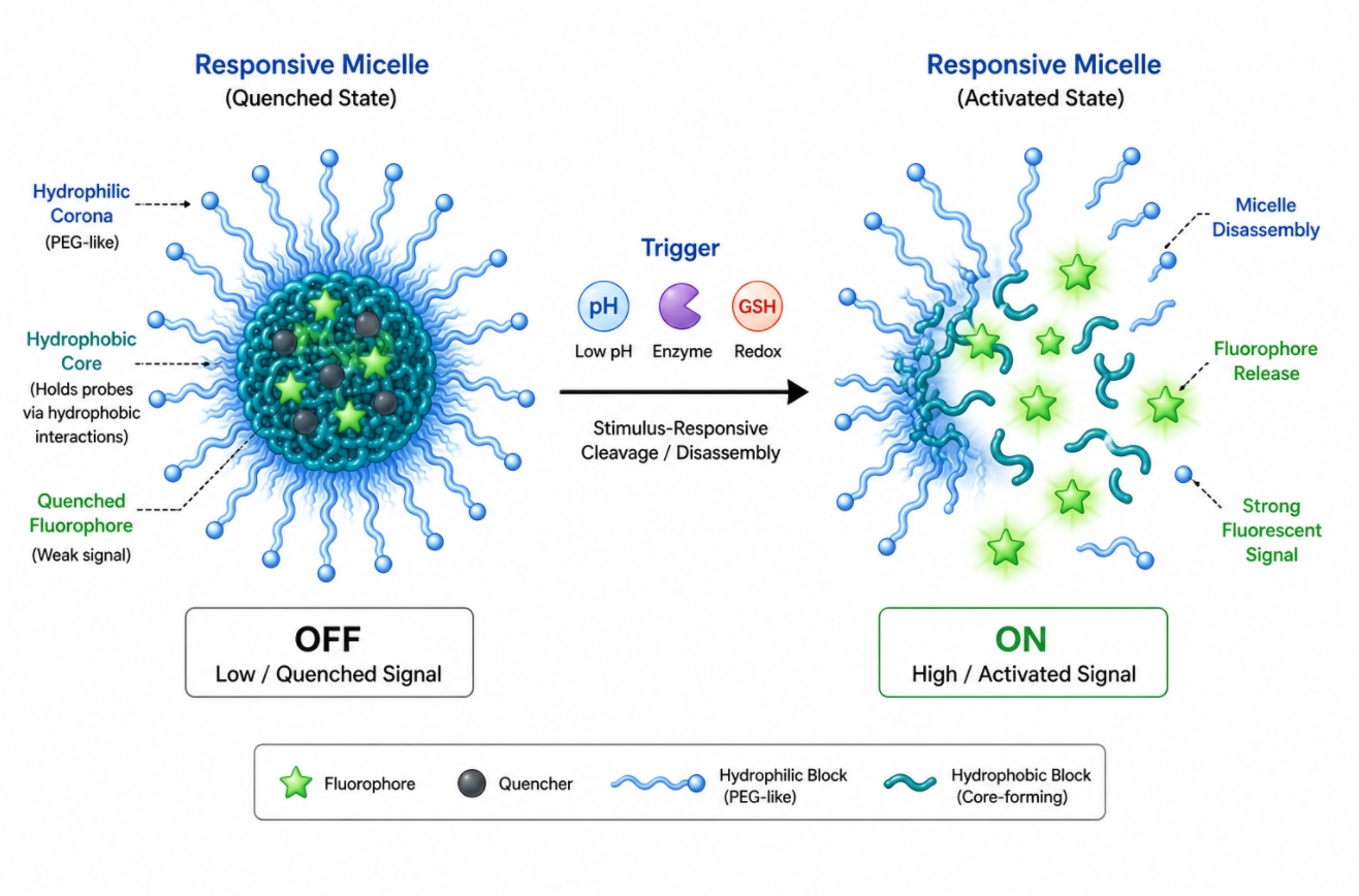 Fig. 2. Responsive polymer micelles enable activatable signal generation in bioimaging (BOC Sciences Authorized).
pH-Responsive Imaging Micelles
pH-responsive micelles are useful when the imaging signal is intended to change in acidic or otherwise altered environments. The response may come from protonation-sensitive probe behavior, micelle destabilization, or cleavage of a pH-labile linkage. In each case, the design challenge is to ensure that the system remains sufficiently stable before the trigger occurs. A pH-responsive imaging micelle that activates too early can lose contrast value, while one that is too stable may show little useful differentiation. The polymer architecture must therefore be tailored so that signal change follows the intended environmental window instead of a broad, poorly selective transition.
Redox- and Enzyme-Responsive Signal Activation
Redox- and enzyme-responsive systems use local chemical changes to activate imaging output or release imaging-related components. A redox-sensitive linkage may alter micelle integrity or probe environment under reducing conditions, while enzyme-responsive motifs can expose or free the imaging unit in the presence of a specific catalytic activity. These approaches are attractive because they link imaging directly to local chemistry rather than simple distribution. However, they require careful control of background stability, since weak selectivity or premature bond cleavage can make the signal difficult to interpret. The best responsive systems are those in which the trigger produces a clear and measurable structural consequence.
AIE and Switch-On Fluorescence Design
Aggregation-induced emission and switch-on fluorescence are especially compatible with polymer micelle design because the assembled nanostructure can regulate molecular motion and packing directly. Instead of being quenched upon confinement, an AIE-active probe may emit more strongly when rotational freedom is restricted inside the micelle or after a defined structural transition. Switch-on fluorescence systems can also be constructed so that probe release, dequenching, or environment-induced change creates a bright signal from a low-background baseline. These strategies are powerful precisely because they make the micelle itself part of the signal mechanism rather than treating it as a neutral container.
Visualizing Cargo Release and Micelle Disassembly
One of the most informative uses of responsive polymer micelles is to visualize cargo release or carrier disassembly indirectly through signal change. In these systems, a fluorescence ratio, intensity shift, or dequenching event may be used to indicate that the micelle has changed state. This can be useful for tracking formulation behavior in real time or for understanding how self-assembly responds to defined environmental conditions. Even so, these imaging claims should be supported by structural characterization and not inferred from signal change alone. A convincing system demonstrates that the imaging readout correlates with actual micelle transformation rather than with unrelated probe redistribution.
Looking for Custom Polymer Micelles for Bioimaging Research?
BOC Sciences supports amphiphilic polymer design, imaging probe integration, responsive micelle development, and characterization workflows for advanced bioimaging applications.
How to Design Polymer Micelles for Multimodal Bioimaging?
Multimodal bioimaging is attractive because no single imaging method can provide every type of information equally well. A multimodal micelle can, in principle, combine complementary signal modes in one platform and improve spatial information, signal confidence, or monitoring capability. But multimodal design is only useful when the structural complexity remains justified by the imaging task. Polymer micelles are helpful here because their core-shell organization allows multiple functional elements to be distributed rationally rather than forced into one overcrowded domain.
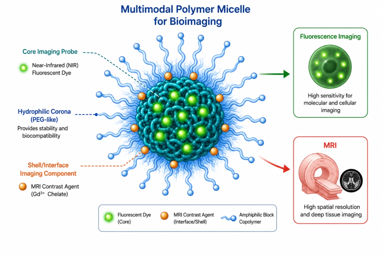 Fig. 3. Multimodal polymer micelles integrate complementary imaging functions in one platform (BOC Sciences Authorized).
Why Combine Two or More Imaging Modalities
The main reason to combine modalities is to overcome the limits of any one signal type. Fluorescence can offer high sensitivity and activatable behavior, while magnetic or other contrast-related strategies may provide different spatial or physicochemical information. A multimodal micelle can therefore support cross-validation of signal origin, improve interpretability, or connect dynamic probe activation with a second more stable imaging output. The key is that the modalities should answer different but complementary questions. If they merely duplicate information, the extra design burden may add little value.
Fluorescence-MRI and Other Common Multimodal Pairings
Fluorescence-MRI pairings are common because they combine an optical readout with a non-optical contrast mode, giving researchers two distinct information channels from the same micelle platform. Other pairings can include photoacoustic-optical or probe-release imaging combined with a second structural signal. The polymer micelle is particularly useful here because one imaging element may be core-loaded while another is attached or associated elsewhere in the assembly. However, such pairings only work if the two signal units do not destabilize each other or distort the self-assembly process that the whole platform depends on.
Balancing Signal Diversity with Structural Simplicity
Every added imaging function increases the demands placed on the polymer architecture. More components mean more competition for space, more opportunities for unfavorable interactions, and more difficulty in maintaining clear structure-property relationships. A good multimodal micelle therefore balances diversity of signal with simplicity of construction. One useful principle is to keep one modality dominant and let the second modality play a supporting role. This often produces a more interpretable system than trying to maximize every signal channel equally within the same micelle.
When Multimodal Design Becomes Unnecessarily Complex
Multimodal design becomes unnecessary when the additional modality does not improve decision-making or when it undermines the colloidal and photophysical stability of the platform. This can happen when probes crowd the micelle, when conjugation density becomes too high, or when the polymer must be modified so extensively that the resulting system is no longer easy to characterize or reproduce. A structurally simple and reliable single-modality micelle may be better than a multifunctional but ambiguous system. The decision to build a multimodal platform should therefore arise from a defined imaging need rather than from a desire to increase complexity.
How to Evaluate Polymer Micelles for Bioimaging Applications?
Evaluation of polymer micelles for bioimaging must connect colloidal characterization with imaging-specific readouts. Standard measurements such as size and stability remain essential, but they are not sufficient. Imaging systems also need to be judged by probe retention, background suppression, signal intensity, activation behavior, and the consistency between structural state and observed signal. A meaningful evaluation strategy therefore asks not only whether the micelle exists, but also whether the imaging output remains reliable as the micelle changes environment or loading state.
Physicochemical Characterization: Size, PDI, CMC, and Stability
Physicochemical measurements provide the baseline for interpreting all later imaging data. Size and polydispersity indicate whether the sample forms a reasonably defined colloidal population. CMC helps estimate how readily the micelle may persist under dilution. Stability measurements reveal whether the structure remains intact or changes in ways that may alter the imaging readout. These parameters do not by themselves prove imaging value, but without them the signal cannot be interpreted confidently. Analytical workflows of this kind can be aligned with polymer characterization services when more formal structure-property evaluation is needed.
Probe Loading, Signal Strength, and Background Control
A useful imaging micelle should show not only measurable probe incorporation but also a favorable balance between signal strength and background. Very high loading can be counterproductive if it causes quenching or instability. Very low loading may preserve signal quality per molecule but fail to generate sufficient total contrast. Background control is equally important, especially for activatable systems intended to remain quiet before stimulation. This means signal evaluation should include more than raw intensity values. It should also ask whether the signal emerges for the right reason and whether unwanted baseline emission or noise remains acceptably low.
Tracking Probe Retention, Leakage, and Activation
Probe retention determines whether the imaging readout genuinely represents the micelle-associated system. Leakage studies help reveal whether the imaging unit remains confined or escapes into the surrounding medium, where it may behave very differently. Activation studies are equally important for responsive systems, because they clarify whether the claimed signal change actually follows the intended trigger. Together, these measurements allow the researcher to distinguish stable imaging micelles from systems that only appear functional at the time of initial preparation. The best formulations are those in which retention and activation data support the same structural interpretation.
Interpreting Multimodal and Responsive Imaging Data
Multimodal and responsive systems require especially careful interpretation because multiple variables change at once. A rise in fluorescence may reflect dequenching, leakage, structural collapse, or intended activation. A second imaging mode may remain stable or may change independently. Proper interpretation therefore depends on combining signal data with structural evidence such as morphology, colloidal stability, or probe localization studies. When the signal channels are consistent with the measured micelle state, the system becomes far more credible. When they are not, the imaging result may be visually impressive but mechanistically weak.
| Evaluation Dimension | Key Question | Representative Readout | Why It Matters | Common Limitation |
|---|
| Colloidal structure | Is the micelle physically well defined? | Size, PDI, CMC, morphology | Establishes a credible structural baseline | Good colloidal data alone do not prove imaging value |
| Probe organization | Is the imaging unit retained and distributed properly? | Loading, retention, leakage studies | Clarifies whether signal belongs to the micelle system | Initial loading may hide later redistribution |
| Signal behavior | Is the observed signal strong and interpretable? | Intensity, background, activation ratio | Connects structure to imaging performance | Signal may be altered by quenching or medium effects |
| Responsive or multimodal performance | Do multiple outputs behave as intended? | Trigger-dependent changes and cross-mode consistency | Supports reliable advanced imaging claims | Complex systems are harder to interpret reproducibly |
Limitations and Design Trade-Offs in Polymer Micelles for Bioimaging
Polymer micelles are highly flexible imaging platforms, but their advantages come with real trade-offs. Increasing signal brightness can undermine structural integrity. Adding more probe can intensify quenching or leakage. Activatable designs can reduce background, yet they may also become less reproducible if the trigger mechanism is too sensitive or poorly controlled. A technically strong resource page should therefore explain not only why micelles are useful, but also where their limits lie and when a different imaging carrier may offer a clearer solution.
Bright Signal vs Stable Micelle Structure
Strong signal often requires a substantial amount of imaging unit within the micelle, but excessive loading can destabilize self-assembly or promote self-quenching. The most emissive system is not always the most structurally reliable. This trade-off is especially important in small micelles where the loading space is limited and core packing is sensitive to disruption. In practice, a slightly lower signal from a stable and well-characterized micelle may be more useful than a brighter but poorly controlled formulation.
High Probe Loading vs Self-Assembly Integrity
Probe molecules are not inert guests. They can compete with polymer packing, alter core polarity, and change the assembled size or dispersity. High loading can therefore undermine the micelle's structural integrity even when total probe incorporation appears impressive. This is particularly relevant for large aromatic imaging units or multifunctional probes that disturb the balance between hydrophilic and hydrophobic segments. Successful bioimaging design usually depends on identifying a loading window that maintains both signal utility and self-assembly reliability.
Responsive Imaging vs Signal Reproducibility
Responsive imaging systems are attractive because they reduce background and make the signal more informative. However, response mechanisms can also introduce variability if small changes in preparation, environment, or probe arrangement alter the activation behavior. A pH-responsive or enzyme-responsive micelle may work well in one set of conditions and behave ambiguously in another. For this reason, responsive designs need stronger validation than constant-output systems. Reproducibility must be demonstrated not only for structure formation, but also for the signal transition itself.
When Another Imaging Carrier May Be More Suitable
Polymer micelles are not ideal for every bioimaging problem. If the imaging unit requires a highly rigid matrix, a permanent solid framework, or extensive inorganic functionality, another carrier type may be more appropriate. In some cases, comparing against broader carrier platform options helps clarify whether micelle self-assembly is truly the best basis for the imaging strategy. The most suitable platform is the one whose structural behavior supports the intended imaging mechanism with the least unnecessary complexity.
Services
Advanced Polymer Synthesis and Formulation Services
At BOC Sciences, we support polymer micelle bioimaging research from the perspective of polymer architecture, probe integration, responsive signal design, and structure-signal evaluation. Our capabilities are oriented toward helping researchers build amphiphilic systems that do more than disperse probes: we help develop micelle platforms in which probe location, self-assembly behavior, functionalization strategy, and imaging output can be tuned together. By combining expertise in polymer synthesis, polymer modification, imaging-oriented micelle preparation, and structure-related analytical assessment, we support more rational development of polymer micelles for optical, responsive, and multimodal bioimaging research.

Amphiphilic Polymer and Block Copolymer Design Support
- Design of amphiphilic polymer systems for stable self-assembly and imaging probe compatibility.
- Adjustment of hydrophilic-hydrophobic balance, molecular weight, and block ratio for micelle formation.
- Selection of suitable polymer segments for optical, responsive, or multimodal imaging tasks.
- Development support for polymer micelle platforms with clear structure-signal logic.
Functionalization and Imaging Probe Integration Support
- Polymer-side functionalization for imaging unit attachment and interfacial control.
- Support for dye incorporation, responsive linker design, and probe organization strategies.
- Integration of imaging-oriented chemistries through side/end-group functionalization.
- Advanced conjugation options through polymer bioconjugation service where needed.
Micelle Preparation and Responsive Imaging Development
- Support for imaging-oriented polymer micelle synthesis and process optimization.
- Optimization of probe loading, size control, colloidal stability, and release-sensitive behavior.
- Guidance on reducing leakage, quenching, and signal instability during formulation design.
- Development pathways for responsive and multimodal micelle systems.
Characterization and Structure-Signal Evaluation Services
- Analytical support for correlating micelle structure with imaging probe behavior.
- Access to morphology-related analysis through polymer structure morphology analysis.
- Evaluation of signal retention, activation, and colloidal performance under defined conditions.
- Support for building more interpretable structure-property datasets in bioimaging research.
Do You Need A Consultation?
BOC Sciences provides tailored support for polymer micelle design, imaging probe integration, responsive formulation optimization, and structure-signal evaluation in bioimaging research.
Products
Unlock New Possibilities with Tailored and High-Performance Polymers
FAQs
Frequently Asked Questions
-
Why are polymer micelles useful for bioimaging instead of free imaging probes?
Polymer micelles are useful because they can improve probe dispersion, protect hydrophobic or aggregation-sensitive imaging units, reduce uncontrolled background, and organize probes within a defined nanoscale environment. Their self-assembled structure also allows imaging output to be linked with probe retention, local activation, or micelle disassembly, which is difficult to achieve with free probes alone.
-
Which imaging modalities are most compatible with polymer micelles?
Fluorescence and near-infrared imaging are among the most naturally compatible modalities because many optical probes benefit from hydrophobic-core loading and controlled molecular packing. Polymer micelles can also support MRI-related, photoacoustic, and multimodal systems, but these usually require more careful control of probe location, loading strategy, and structure-signal relationships than simple optical formulations.
-
How does probe location affect polymer micelle imaging performance?
Probe location strongly affects whether a polymer micelle produces stable and interpretable imaging data. Core-loaded probes may be better protected but more vulnerable to overpacking and quenching. Corona-associated probes may be more exposed and responsive but less shielded from interference. The chosen location determines leakage risk, background behavior, signal mechanism, and how reliably imaging reflects micelle structure.
-
Are responsive polymer micelles important in bioimaging?
Responsive polymer micelles are important when the goal is to obtain activatable or low-background imaging rather than constant signal output. By coupling signal generation to pH change, redox conditions, enzymatic activity, or micelle disassembly, these systems can make imaging more informative. Their value depends on achieving selective activation without sacrificing baseline colloidal stability and signal reproducibility.
-
When should multimodal imaging be considered for polymer micelles?
Multimodal imaging should be considered when one signal type alone cannot provide enough information about distribution, activation, or structural behavior. Polymer micelles are suitable when their compartmentalized architecture can support two complementary imaging modes without excessive interference. Multimodal design is less useful when the second modality adds complexity but does not improve interpretation or decision-making meaningfully.
-
What are the main limitations of polymer micelles for bioimaging?
The main limitations include probe leakage, aggregation-related quenching, restricted loading windows, and increasing complexity in responsive or multimodal systems. Because imaging output depends strongly on probe organization, even small structural variations can alter signal behavior. Polymer micelles are most effective when the imaging mechanism matches the self-assembled architecture rather than forcing unsuitable probes into unstable colloidal systems.
